NWO Roadmap NL-BioImaging: A Dutch infrastructure of advanced microscopy
 Posted by MarkHink in News
  Comments Off on NWO Roadmap NL-BioImaging: A Dutch infrastructure of advanced microscopy
 20
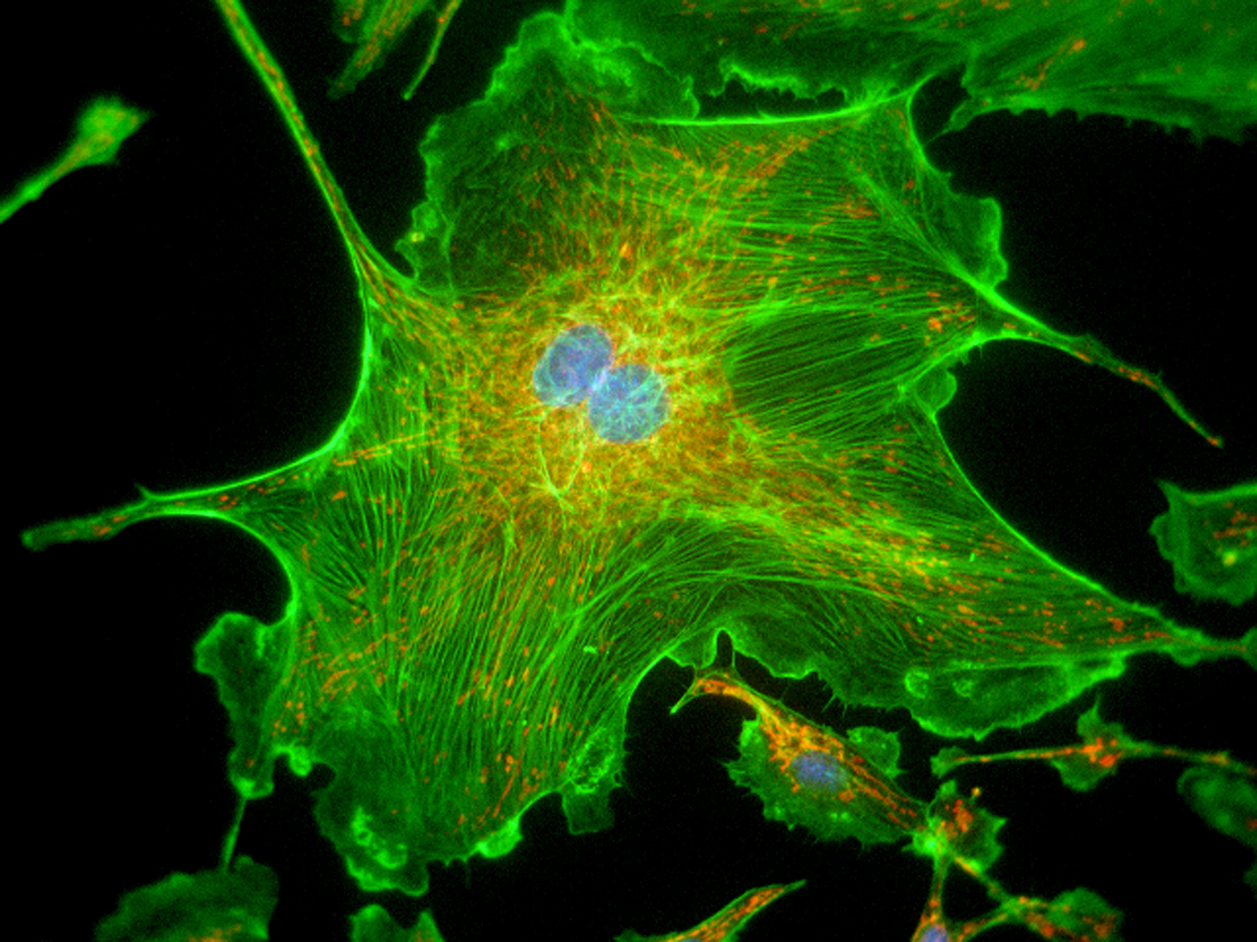
Advanced microscopy to understand life and fight disease.
NL-BI is the Dutch infrastructure network for advanced light microscopy in life sciences integrated in the ESFRI program Euro-BioImaging and closely linked to the Dutch Society for Microscopy (NVvM). Participants are all Dutch universities, UMCs, as well as thematic research institutes. .
NL-BI will develop and connect
state-of-the-art technologies and services for functional imaging of live
processes to investigate phenotypic heterogeneity, responses, and mechanisms. A
dedicated national FAIR data-management and analysis team will link all Dutch
microscopy facilities, and enable greater reuse, mining, and interlinking of
large amounts of image data generated using novel AI tools. High-content and
functional imaging will create the necessary fundaments to integrate microscopy
with the omics technologies (‘visual omics’), requiring on-the-fly data
analysis and smart microscopy. By connecting these components, NL-BI will
enable fundamental insights and innovative applications, such as development of
biomarkers, vaccines, and personalized medicine for complex disorders as
cardiovascular disease, cancer, diabetes, and neurodegenerative disorders.
Key aims of national relevance are to further develop advanced microscopy, create the necessary infrastructure, and provide expertise to enable the Dutch research community to:
- Use new molecular probes and biosensors for functional imaging
- Apply fast 3D imaging technologies for functional imaging in complex live-cell systems
- Carry out high-content microscopy screens for cell phenotype changes
- Bridge scales towards electron microscopy and intravital microscopy
- Automate complex microscopy workflows and on-the-fly data analysis to massively increase experimental throughput and provide unbiased, researcher-independent output
- Use a national network for FAIR image data management providing infrastructure, training, and easy-to-use workflows for data analysis of biological images by state-of-the-art neural networks
- Establish a nation-wide training and outreach platform for scientists at all levels
By providing coordinated access to the Netherlands’ best imaging technology and analysis platforms, the NL-BI roadmap proposal will achieve much-needed excellence in advanced optical microscopy to secure the international competitiveness of Dutch life science research.
LCAM: New state-of-the-art equipment
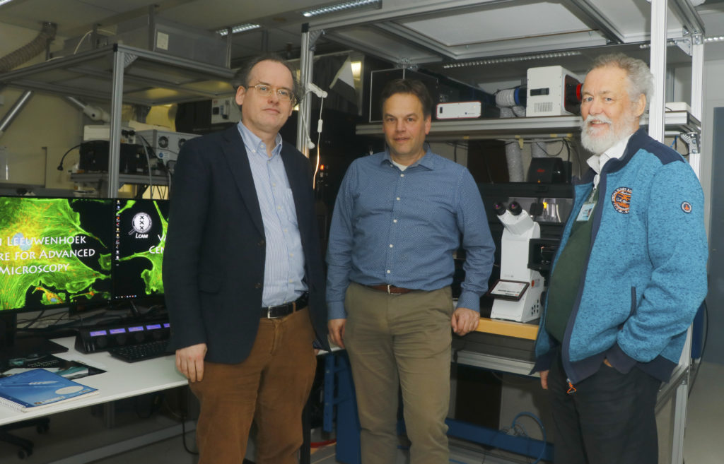
With the financing of the NL-BioImaging Roadmap, all participating Dutch microscopy centres can bring their infrastructure to the top level, specific techniques and expertise are connected, and a national network for data analysis and management will be set up. This opens the door to new scientific breakthroughs by thousands of life scientists in the Netherlands. In LCAM Amsterdam, for example, 4 million will be invested in equipment and 1.5 million in support, which gives an enormous boost to state-of-the-art microscopy cell biology. At the University of Amsterdam, under supervision of Dorus Gadella (LCAM-UvA and Swammerdam Institute for Life Sciences), investments will be made in a new super-resolution microscope, which produces 3-5 times sharper images than conventional microscopes by making smart use of shortening of the (nanosecond 10-9 s) fluorescence lifetime. Gadella: ‘We were able to test this concept with new switchable fluorescent proteins and biosensors developed by ourselves, so we know this technique will actually work in living cells and yield dynamic super-resolution information.’
Consortium leader Eric Reits (LCAM – Amsterdam UMC) will install a high-content 3D microscope with which fast processes with fluorescent biosensors in organoid models can be analysed on-the-fly. Reits: ‘Using self-steering microscopy scripts, the microscope is fast enough to quantify the effects of potential therapeutic strategies and to isolate cells with an interesting phenotype for proteome, metabolome and genome analysis, i.e. visual omics.’ Kees Jalink (LCAM-NKI) invests in the development of automated pooled microscopy screening, in which extensive compounds and CRISPR knockout libraries are tested in cells for changes in phenotype. Jalink: ‘By using different fluorescent biosensors that we develop at LCAM, researchers can screen which compounds and genes effectively change a phenotype in cells.’
Education
All these developments put LCAM and Amsterdam further on the map. Since Gadella and Reits coordinate the unique biomedical Master’s track Cell Biology and Advanced Microscopy, in collaboration with Jalink, the new technologies and analysis scripts will also be used immediately in education, thus training the new generation of cell biologists. Reits: ‘Within this master’s program, students learn hands-on how to make optimal use of the various microscopes and they learn about the possibilities of AI data analysis. Combined with internships at (inter)national microscopy expertise groups, they are well prepared to immediately implement these state-of-the-art techniques in biomedical research as PhD students.’
LCAM centre among best in Europe
15 December 2015
The van Leeuwenhoek Centre for Advanced Microscopy (LCAM), a collaboration of the Netherlands Cancer Institute, the AMC-UvA and the UvA’s Faculty of Science, is according to Euro-BioImaging one of the leading microscopy centres in Europe. The centre has therefore been officially endorsed as a node of this European consortium.
Euro-BioImaging is an emerging European partnership seeking to use Europe’s wide-ranging bioimaging expertise as efficiently as possible. With this quality seal, LCAM has attained the highest qualification in the development and application of advanced microscopy and imaging analysis and the provision of technology to users. A total of 28 European microscopy centres were declared nodes.
According to director Dorus Gadella, ‘It is Europe’s only designated “flagship node” in functional imaging microscopy.’ He explains: ‘functional imaging is the direct visualization and quantification of molecular processes in living cells with fluorescent biosensors. For instance, the interaction, movement and conformation changes of biomolecules and structures in cells. Or how external signals are processed by cells and how DNA is transcribed in the cell nucleus.’
Euro-BioImaging consortium
The declaration of the 28 European microscopy centres is a momentous step in the realisation of the European Strategy Forum on Research Infrastructure (ESFRI) Euro-BioImaging Roadmap. The ESFRI is a strategic instrument to develop the scientific integration of Europe and to strengthen its international outreach. The Euro-BioImaging consortium aims to build an infrastructure of wide-ranging, state-of-the-art imaging technologies. Thanks to its open access structure, it will in future provide life scientists in Europe and beyond with access to sophisticated facilities for biological, molecular and medical research.
Microscopy in the Netherlands
ESFRI quality seals have been awarded to nine centres in the Netherlands, placing our country, the technique’s birthplace, among Europe’s bioimaging leaders. With this European recognition, the prominence of the Dutch research tradition in this field has been confirmed. The Dutch centres are united in the NL BioImaging Advance Microscopy roadmap, coordinated by the University of Amsterdam.
LCAM Advanced Microscopy Course
LCAM Advanced Microscopy Course
June 23-27, 2014
This course is organized by the Leeuwenhoek Centre for Advanced Microscopy (LCAM) and provides an in-depth view of specific advanced imaging techniques used at imaging centers of AMC, NKI and FNWI. The course focuses fully on intensive hands-on practical sessions and interactive discussions with experts in the various microscope techniques. On the final day participants present their own research topic and discuss which techniques should be implemented in their project.
During the course participants will visit the imaging centres of three institutes in Amsterdam, the NKI (Dutch Cancer Institute), the FNWI (UvA Science Park) and the AMC. In each institute the part will focus in depth on specific advanced imaging techniques.
Techniques:
– Measuring intra- and inter-molecular protein interactions using FRET and FLIM
– Confocal microscopy methods including spinning disk (CLSM)
– Superresolution microscopy
– Confocal quality control
– Correlative microscopy (bridging fluorescence microscopy and electron microscopy)
– Usage of fluorophores such as FlAsH/ReAsH
Target audience:
PhD students and postdocs. An introduction course like the Basic Microscopy course is a prerequisite for the Advanced Microscopy Course.
Course organizers:
AMC Ron Hoebe, Eric Reits, Jan Stap, Henk van Veen
FNWI Dorus Gadella, Joachim Goedhart, Mark Hink, Erik Manders
NKI Lenny Brocks, Kees Jalink, Lauran Oomen
More information and application:
From Eric Reits / e.a.reits@amc.uva.nl / tel. +31 (0)20 566 6259
LCAM Website restyled
Today the LCAM website got restyled. Please let me know what you think about the new look!
LCAM
News
- NWO Roadmap NL-BioImaging: A Dutch infrastructure of advanced microscopy
- LCAM centre among best in Europe
- LCAM Advanced Microscopy Course
- LCAM Website restyled